Diabetic retinopathy can damage the retina for years before it blurs a single word on a page. Many adults first learn they have it during a routine dilated exam, long after the earliest changes began.
After age 50, the odds of developing diabetes and several other retinal conditions climb, and most of them start without pain or obvious vision loss. November is Diabetic Eye Disease Awareness Month, a good reminder to schedule a retinal exam.
Keep reading to learn more about how your retina changes with age, which conditions become more common, and how often you should be seen.
What Changes in Your Retina After 50?
The retina is the thin, light-sensitive tissue lining the back of the eye, and it ages along with the rest of your body. The vitreous is the clear gel that fills the eye, and as it shrinks with age, it can tug on the retina as it pulls away. Small blood vessels become less flexible, and the macula, the center of the retina responsible for sharp detail, becomes more vulnerable to wear.
None of these changes guarantee a problem, but together they raise the baseline risk for several retinal diseases. Many people also start needing reading glasses around this age, but that change happens in the lens at the front of the eye. The hardest part of catching retinal diseases is that they often don’t cause symptoms until vision is permanently lost.
Why Diabetes Raises the Stakes
Type 2 diabetes becomes more common with age, and the risk of eye complications grows the longer a person lives with it. Someone diagnosed in their 50s may have had elevated blood sugar for several years before the diagnosis, which means retinal changes can begin earlier than expected.
Over time, high blood sugar can damage the blood vessels in your retina, weakening the walls of tiny capillaries until they swell, leak, or close off. This process is called diabetic retinopathy, and it typically moves through stages:
- Mild nonproliferative retinopathy: Small bulges called microaneurysms form in the capillary walls. Vision is usually normal.
- Moderate to severe nonproliferative retinopathy: More vessels become blocked, and parts of the retina start losing their blood supply.
- Proliferative diabetic retinopathy: The eye grows new, fragile blood vessels that can bleed into the vitreous or pull on the retina.
At any stage, fluid can leak into the macula and cause diabetic macular edema. This is one of the most common reasons people with diabetes notice blurry central vision. Because the early stages usually cause no symptoms, a person may feel their eyesight is fine while damage builds. Diabetic retinopathy cannot be reversed, but treatment at the right time can slow or stop further vision loss, and catching it early gives your eye doctor more options.
Blood pressure and cholesterol also tend to rise with age, and both add stress to the same small vessels that diabetes affects.
Other Retinal Conditions That Become More Common With Age

Diabetes is only one part of the picture when it comes to age-related retinal conditions. Age-related macular degeneration is a leading cause of central vision loss in older adults. The dry form develops slowly over years, but the wet form involves abnormal blood vessels beneath the macula and can affect vision within weeks.
As with many retinal diseases, early macular degeneration often has no symptoms. Some people first notice it when straight lines look wavy, or a blurry spot appears in the center of their vision.
A retinal vein occlusion happens when a vein carrying blood away from the retina becomes blocked. It is more common in people with high blood pressure, diabetes, or glaucoma.
A shrinking vitreous can also cause a retinal tear, which may lead to retinal detachment if left untreated. A single dilated retinal exam lets your eye doctor check for all of these conditions at once.
What Happens During a Dilated Retinal Exam?
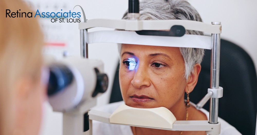
A dilated eye exam is the most direct way to see the full retina. Your eye doctor places drops in your eyes to widen the pupils and waits about 20 to 30 minutes for them to take effect. Then your eye doctor examines the retina, macula, optic nerve, and blood vessels with specialized lenses and lights.
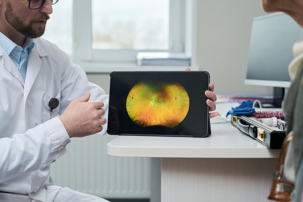
Many visits also include imaging such as optical coherence tomography (OCT). This scan produces cross-section images of the retina’s layers and can reveal swelling in the macula before you notice any blur. Fundus photography captures detailed pictures of the back of the eye, giving your eye doctor a baseline to compare against at future visits.
How Often Should You Be Seen?
Most people with diabetes need a dilated eye exam at least once a year, and some need more frequent visits once retinopathy is found. Your eye doctor may also shorten that interval if your blood sugar has been difficult to control or if you already show signs of macular edema. Adults over 50 without diabetes should follow the schedule their eye doctor recommends based on personal risk factors and family history.
Between visits, contact an eye doctor right away if you notice any of the following:
- A sudden shower of new floaters or flashes of light
- A shadow or curtain moving across part of your vision
- Straight lines that look bent or wavy
- Sudden blurring or loss of vision in one eye
Specialized Retina Care in St. Louis
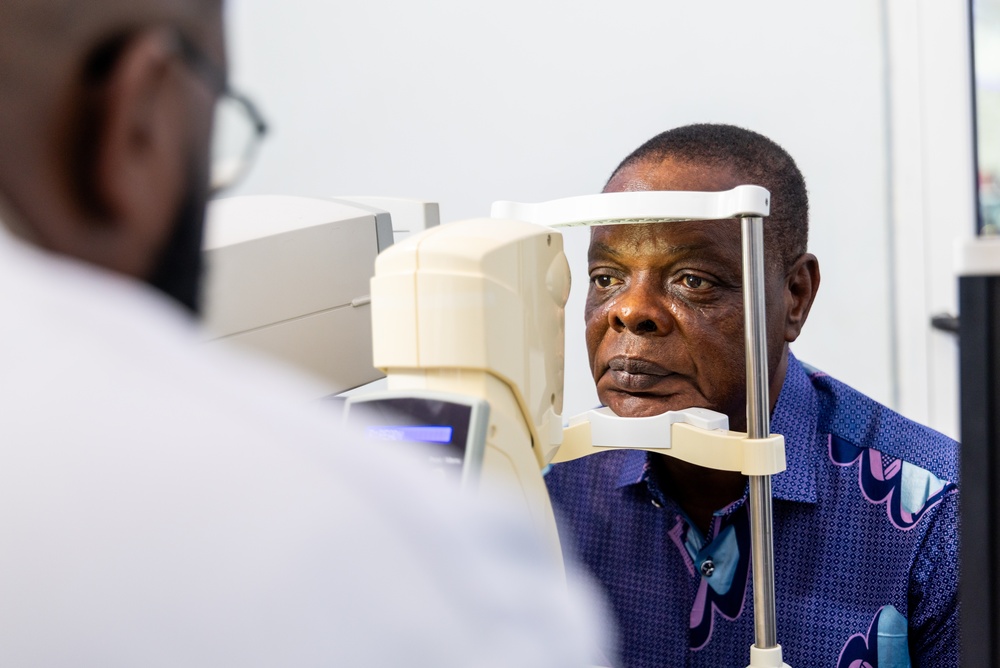
The board-certified retina specialists at Retina Associates of St. Louis focus on retinal and vitreous diseases every day. That focus allows your eye doctor to recognize subtle diabetic changes early and track them over time with advanced imaging.
When monitoring alone is no longer enough, your eye doctor can recommend retinal treatments such as anti-VEGF injections, laser therapy, or vitrectomy. For many patients, the most valuable part of care is a consistent exam schedule that catches changes while vision is still strong.
Schedule an appointment at Retina Associates of St. Louis in St. Louis, MO, to have your retina examined and catch changes before they affect your vision.